Today we’ll be looking at different types of medical imaging tests, as listed in a UVA Radiology article.
X-Rays
These are simple, painless tests that use ionizing radiation to produce images of the structures or bones in your body. These are used to diagnose fractures, arthritis, osteoporosis, infections, swallowed items, breast cancer, even digestive tract problems. It’s not an extended test either: only 10-15 minutes where you lie, sit, or stand while the machine takes the images. To get the best results, you could be asked to move to different positions for different angles of the x-rays.
CT Scans
This particular test uses x-rays to create a clearer picture of the inside of your body. This includes bones, blood vessels, and soft tissues. It’s another test that uses ionizing radiation, which also doesn’t take long. It’s used to diagnose traumatic injuries, fractures, tumors, cancers, vascular disease, heart disease, infections, and can even be used to guide biopsies. You lie on a table that moves you into a giant doughnut-shaped scanner. The tube is a big x-ray machine that rotates around you and takes images.
MRI
This is one of the more extended tests. MRIs use magnetic fields and radio waves to create a more detailed image of the organs and tissues in your body. The duration is around 45 minutes to an hour, and it’s used to diagnose Multiple Sclerosis, aneurysms, stroke, spinal cord disorders, tumors, blood vessel issues, or even joint or tendon injuries. You have to lie on a table that slides you into the MRI machine that’s deeper and narrower than a CT scanner, but it still looks like a giant donut. The MRI machine’s magnets then create loud tapping or thumping noises during the procedure while it takes photos.
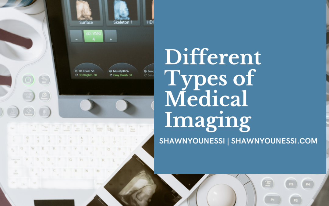
Ultrasound
This test uses high-frequency sound waves to take pictures of the critical organs and structures of your body. It can be shorter than an MRI but longer than an X-ray. A technician first applies gel to your skin and then presses a probe against it and then moves the probe to capture essential images from the inside of your body. This is used to diagnose gallbladder disease, breast lumps, genital/prostate issues, joint inflammation, blood flow problems, to guide biopsies, though it’s most commonly used during pregnancy.